Chromosome diseases.
Date: 2015-10-07; view: 918.
Hereditary diseases of the human being.
THE BASIC THEORETICAL ITEMS OF INFORMATION
About 5 % of human population has hereditary disorders. However in most cases these are not heavy forms and the people are practically healthy. Only less that one percent of population has bad hereditary diseases. But the diseases are the causes of invalidism and death.
Hereditary diseases of the human being are diseases that are caused by mutations (changes within genotype).
Hereditary diseases are different from congenital diseases. Congenital diseases are diseases that are manifested just after birth.
Categorization (classification) of hereditary diseases
1. Chromosome diseases:
a) Chromosome diseases are caused by changing of number and shape of autosome chromosomes.
b) Chromosome diseases are caused by changing of sex chromosomes' number.
2. Gene diseases (molecular diseases).
a) Plural congenital disorders of ontogenesis.
b) Diseases of metabolism disorders.
3. Multifactorial disorders.
Chromosomal diseases caused by changing of number or shape of chromosomes.
Commonest chromosomal disorders seen in newborns
| Disorder | Birth frequency |
| Balanced translocation | 1 in 500 |
| Unbalanced translocation | 1 in 2000 |
| Pericentric inversion | 1 in 100 |
| Trisomy 21 | 1 in 700 |
| Trisomy 18 | 1 in 3000 |
| Trisomy 13 | 1 in 5000 |
| 47,XXY | 1 in 1000 males |
| 47,XYY | 1 in 1000 males |
| 47,XXX | 1 in 1000 females |
| 45,X | 1 in 5000 females |
The most distributed chromosomal diseases caused by changing of number and shape of autosome chromosomes are Down Syndrome (trisomy 21, Down's syndrome, mongolism), Edwards' syndrome (trisomy 18), Patau's syndrome (trisomy 13), and cat cry syndrome (cri du chat, Lejeune's syndrome).
For Down's syndrome the karyotypes 47,21+ (simple trisomy 21), 47,21+/46 (mosaic variant), or 46,Т21/13; 46,Т21/14; and 46,Т21/15 (translocation variant) are typical. In all cases a person has superfluous 21-th chromosome.
Simple trisomy means each cell of organism have a superfluous 21-th chromosome. Mosaic variant means the part of organism's cells has a superfluous 21-th chromosome, but another part doesn't have. Translocation variant means each cell of organism has a superfluous 21-th chromosome, but the chromosome is translocated on the non-homologous chromosome (for instance on 13-th, 14-th, or 15-th).
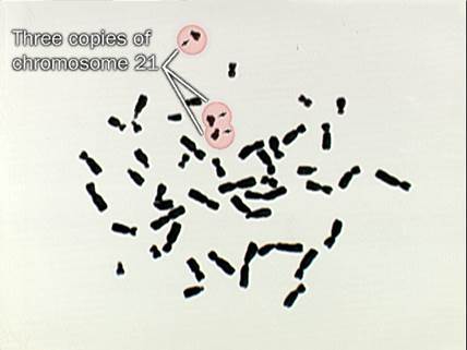
Figure 31.1 Down's syndrome. Karyotypes 47,21+ (simple trisomy 21),
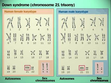
Figure 31.2 Down's syndrome. Idiogram (karyogram) 47,21+ (simple trisomy 21),
Clinical presentation of Down's syndrome include by lag of physical growth and development, by lag of mental and sexual development. There are some malformations. Life interval may by 30-40 years. But in some cases they may die more early.
Clinical presentation of Down's syndrome is on the figure 31.3.
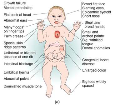
Figure 31.3. Clinical presentation of Down's syndrome.
Edwards' syndrome typical karyotypes (figure 31.4) are 47,18+ (trisomy 18), and 47,18+/46 (mosaic variant).
Definition.Edwards' syndrome is caused by an extra copy of chromosome 18. For this reason, it is also called trisomy 18 syndrome. The extra chromosome is lethal for most babies born with this condition. It causes major physical abnormalities and severe mental retardation, and very few children afflicted with this disease survive beyond a year.
In the case of Edwards' syndrome, the child inherits three, rather than two, copies of chromosome 18. Trisomy 18 occurs in approximately one in every 3,000 newborns and affects girls more often than boys. Women older than their early thirties have a greater risk of conceiving a child with trisomy 18, but it can occur in younger women.
Causes and symptoms.A third copy of chromosome 18 causes numerous abnormalities. Most children born with Edwards' syndrome appear weak and fragile, and they are often underweight. The head is unusually small and the back of the head is prominent. The ears are malformed and low-set, and the mouth and jaw are small. The baby may also have a cleft lip or cleft palate. Frequently, the hands are clenched into fists, and the index finger overlaps the other fingers. The child may have clubfeet and toes may be webbed or fused.
Numerous problems involving the internal organs may be present. Abnormalities often occur in the lungs and diaphragm (the muscle that controls breathing), and heart defects and blood vessel malformations are common. The child may also have malformed kidneys and abnormalities of the urogenital system.
Diagnosis.Physical abnormalities point to Edwards' syndrome, but definitive diagnosis relies on karyotyping. Karyotyping involves drawing the baby's blood or bone marrow for a microscopic examination of the chromosomes. Using special stains and microscopy, individual chromosomes are identified, and the presence of an extra chromosome 18 is revealed.
Trisomy 18 can be detected before birth. If a pregnant woman is older than 35, has a family history of genetic abnormalities, has previously conceived a child with a genetic abnormality, or has suffered earlier miscarriages, she may undergo tests to determine whether her child carries genetic abnormalities. Potential tests include maternal serum analysis or screening, ultrasonography, amniocentesis, and chorionic villus sampling.
Treatment.There is no cure for Edwards' syndrome. Since trisomy 18 babies frequently have major physical abnormalities, doctors and parents face difficult choices regarding treatment. Abnormalities can be treated to a certain degree with surgery, but extreme invasive procedures may not be in the best interests of an infant whose lifespan is measured in days or weeks. Medical therapy often consists of supportive care with the goal of making the infant comfortable, rather than prolonging life.
Prognosis.Most children born with trisomy 18 die within their first year of life. The average lifespan is less than two months for 50% of the children, and 90-95% die before their first birthday. The 5-10% of children who survive their first year are severely mentally retarded. They need support to walk, and learning is limited. Verbal communication is also limited, but they can learn to recognize and interact with others. Prevention.Edwards' syndrome cannot be prevented.
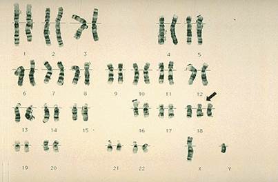
Figure 31.4 Edwards' syndrome typical karyotypes (47,18+)
Patau's syndrome typical karyotypes are 47,13+ (trisomy 13), and 47,13+/46 (mosaic variant). Clinical presentation of Patau's syndrome includes some hard malformations. Life interval is about one years or less. As a rule they dies during neonatal period.
The typical karyotypes of cat cry syndrome is 46,5p-. It means that the cause of the syndrome genesis is deletion of short haul of 5-th chromosome. Clinical presentation of cat cry syndrome includes some hard malformations. One of main symptoms is disorder of vocal apparatus structure. Therefore the newborn emits sound like the cat's meow. Sick children have been living less than one year.
The classical examples of hereditary chromosome disease that are caused by sex's chromosomes number change are Klinefelter's syndrome, Turner's syndrome (gonadal dysgenesis), polysomy (polysomia) X chromosome syndrome, polysomy Y chromosome syndrome.
Turner's syndrome typical karyotypes (figure 31.5) is 45,X (or 45,X0). Clinical presentation ofTurner's syndrome is at the figure 35.6. There is woman's phenotype, intellectual growth and development is normal, but there is sexual and physical growth underdevelopment. Distinguishing feature of the syndrome is fold of skin on the neck.
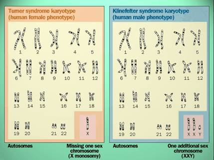
Figure 31.5 Typical idiogram of Klinefelter's syndrome and Turner's syndrome
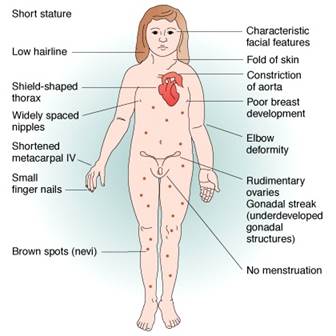
Figure 35.6. Clinical presentation of Turner's syndrome
Polysomy X chromosome syndrome is characterized by karyotype 47,ХХХ (trisomy X), 48,ХХХХ (tetrasomy X), 49,ХХХХХ (pentasomy). When the karyotype 46,ХХХ is present the clinical presentation is following: woman's phenotype, secondary sex characteristics are good developed, there is insignificant mental retardation and insignificant euphoria. In the case of karyotypes such as 48,ХХХХ and, 49,ХХХХХ there are severe mental retardation, developmental lags.
Klinefelter's syndrome is characterized by karyotype (figure 31.5) 47,ХХУ, or 48,ХХХУ (more seldom). Phenotype is man's, but the sex characteristics are manifested a little. Intellect is normal. Function of reproduction is lost. Clinical symptoms of the syndrome are presented at figure 31.7.
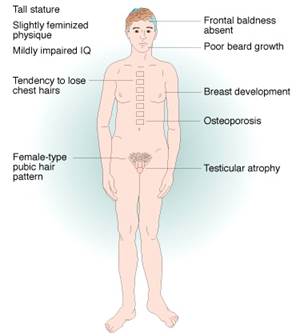
Figure 31.7. Clinical symptoms of Klinefelter's syndrome
Polysomy Y chromosome syndrome is characterized by karyotype 47,ХУУ, of, more seldom, 48,ХУУУ, 49,ХУУУУ. Man's phenotype, very good physical development, the body's length more then 190 centimeters is presence. Different degrees a backlog in the mental development may be present too. The aggressiveness is distinctive.
| <== previous lecture | | | next lecture ==> |
| CARRYING OUT of the CLOSING TEST CONTROL | | | Gene diseases (molecular diseases). |